- Charcot
-
by tmi
From the Archive: This post was originally published on October 22, 2010. It has been republished as part of our ongoing commitment to sharing evidence-based information on diabetic foot care.
Charcot Foot Deformities: O ver the last decade or so, it seems as though the world diabetes community has awakened to the importance of Charcot Foot deformities in this population. Since it’s description by Jean-Martin Charcot himself in 1868, this entity has fascinated many of us who are often perplexed not just by its pathophysiology, but its presentation and management as well. Although Charcot, a leading physician of his time who practiced at La Salpetrierre in Paris (“The Grand Asylum of Human Misery”) described the arthropathy of locomotor ataxia in patients with tertiary syphilis, we now recognize this disorder to be most commonly associated with diabetic peripheral neuropathy. Although his first descriptions of this arthropathy dealt with joint disorders of the knee, Charcot also was the first to publish its more common involvement in the joints of the foot. In fact, he coined the term “Pied Tabetique” in his 1883 clinicopathologic description of this deformity. Figure 1. Is copied from his 1883 paper and illustrates an “exploded” model of the skeleton of what we now call a “Charcot foot” with primarily midfoot involvement.
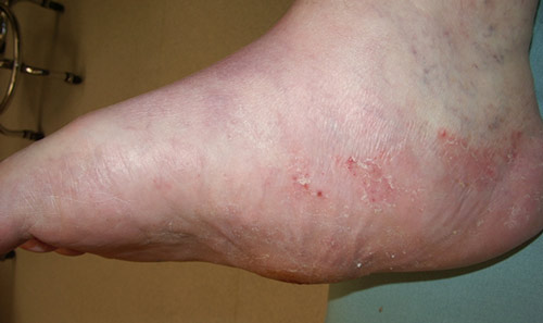
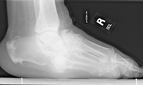
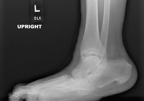
A typical Charcot foot deformity that most of us would easily recognize is presented in Figures 2 and 3. Any patient who presents with long standing diabetes (10 or more years), established sensory neuropathy, with or without a history of injury, and a warm, unilateral swollen foot with such deformity should be considered to have neuropathic arthropathy until proven otherwise. Early diagnosis is critical to prevent the natural progression of the deformity.
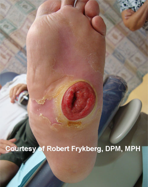
When a midfoot ulcer is present as well, we must also be concerned about concomitant infection and underlying osteomyelitis. (See FootNote #11, July 2009). Presuming there is no ulceration or infection present, the management primarily consists of immobilization and non-weightbearing(NWB) in a total contact cast or removable cast walker. Although several studies have indicated that weight bearing might have no adverse consequences, it is always safer to arrest the destructive cycle by avoiding stress through the foot. The period of NWB will generally be three or four months depending on the degree of deformity present at diagnosis. Following resolution of edema and inflammation (assessed by clinical examination and dermal temperature measurements), a gradual resumption of protected partial weight bearing may commence in the aforementioned removable cast walker/walking brace/ or Charcot Restrained Orthotic Walker (CROW) brace. The goal is to preventfurther deformity while converting the acute Charcot foot into a chronic/quiescent condition. A plantigrade deformity is ideal since this can fairly easily be shod in a therapeutic or custom molded shoe. When deformity has progressed to a “rocker-bottom” configuration (see Figures 4a and 4b below), the treatment becomes more complex, since recurrent ulceration often ensues. Surgery then often becomes necessary to re-establish a plantigrade foot or to reconstruct the arch. Surgery is also indicated for severe, unstable deformities (often involving the ankle or hindfoot) that cannot be successfully treated by bracing and footwear alone. While once considered ill-advised, surgery for the complicated Charcot deformity has become more commonplace in recent years. (Refer to the recent ezine and blog that I commented on. Jarrod’s Practice Perfect) Nonetheless, patients need to be advised that such surgeries are complicated and often require a lengthy convalescence. Intercurrent infection can also place the limb at risk for amputation.
Unfortunately, at this time we do not have any level 1 evidence to guide us pertaining to those patients (or deformities) best treated by surgery and those best suited to conservative management. Generally, however, most practitioners will attempt to treat all deformities conservatively at first. If conservative management fails to prevent recurrent ulceration or if the foot deformity is not amenable to custom footwear management, it is indeed prudent to consider surgical options, depending upon the patients’ concurrent medical comorbidities. Best regards,
REFERENCES
|